SegR3D
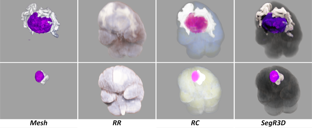
SegR3D: A multi-target 3D visualization system for
realistic volume rendering of meningiomas

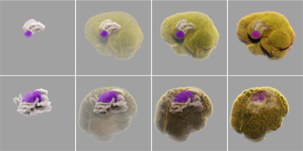
importance transfer function – regulates the visual appearance.


User interaction video
Changing observation angle/adjusting importance transfer function/using real-time denoising
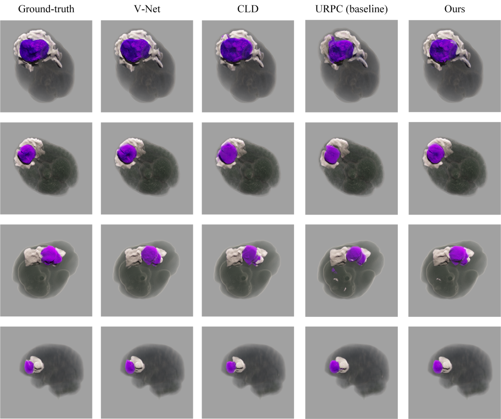
High-precision semi-supervised segmentation

User Study
Descriptions

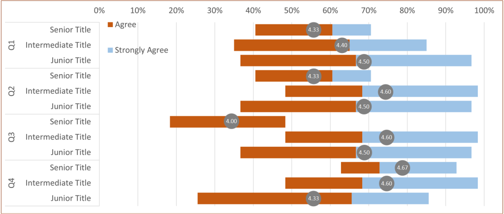
| · Q1: The results of SegR3D are particularly beneficial for comprehending the location and distribution of tumors and SNFH within the intracranial space. |
| · Q2: Our visualization outcomes significantly aid in understanding the shape and size of tumors and SNFH. |
| · Q3: SegR3D is the optimal tool among these methods for tumor analysis. |

| · Q4: The current precision of the SegR3D system is sufficient for tumor analysis and surgical planning purposes. |
Rating Results

Other opinions and suggestions
(1) Is it possible to visualize the relationship between tumors and blood vessels? Attention must be given to tumors closely associated with blood vessels during surgery, as neglecting to protect these vessels can lead to substantial intraoperative bleeding and postoperative complications.
(2) Can the relationship between tumors and the skull be demonstrated? For tumors located in specific regions, such as the skull base, it is crucial to examine the relationship between the tumor and adjacent bone for guidance in surgical procedures.
(3) Is it feasible to illustrate the relationship between tumors and nerves? Tumors closely intertwined with nerves require careful protection during surgery, as damaging the nerves can result in postoperative functional impairments corresponding to the affected neural pathways.
(4) Can users selectively display specific brain tissues (e.g., only showing the frontal lobe and lesions)?